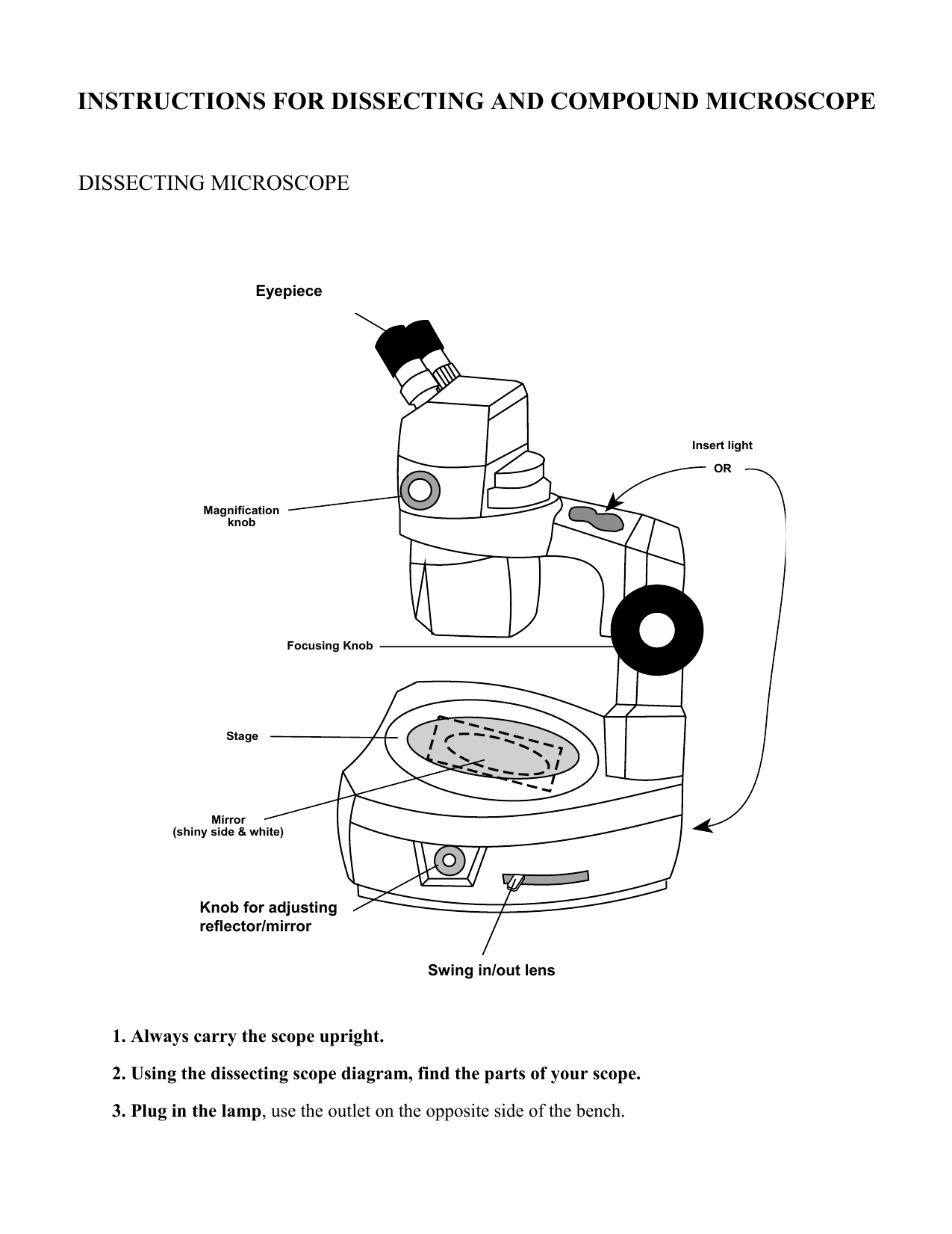
38 dissecting microscope diagram with labels
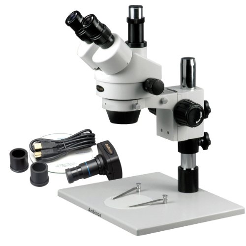
Dissecting microscope (Stereo or stereoscopic microscope)- Definition ... Stereo Zoom Dissecting Microscope-They are trinocular or binocular dissecting microscopes with a zooming range of 6.7x-45x. They can be attached to the digital camera which takes photos of the viewing images. They have a dual-LED illuminator and they can rotate at 360°. Microscope Types (with labeled diagrams) and Functions These microscopes work on the principle called contrast-enhancing technique that is utilized to produce high-contrast images to view them with more accuracy and clarity. Phase-contrast microscope labeled diagram Phase-contrast microscope functions: Its applications areas include In cases where the specimen is colorless and is very tiny
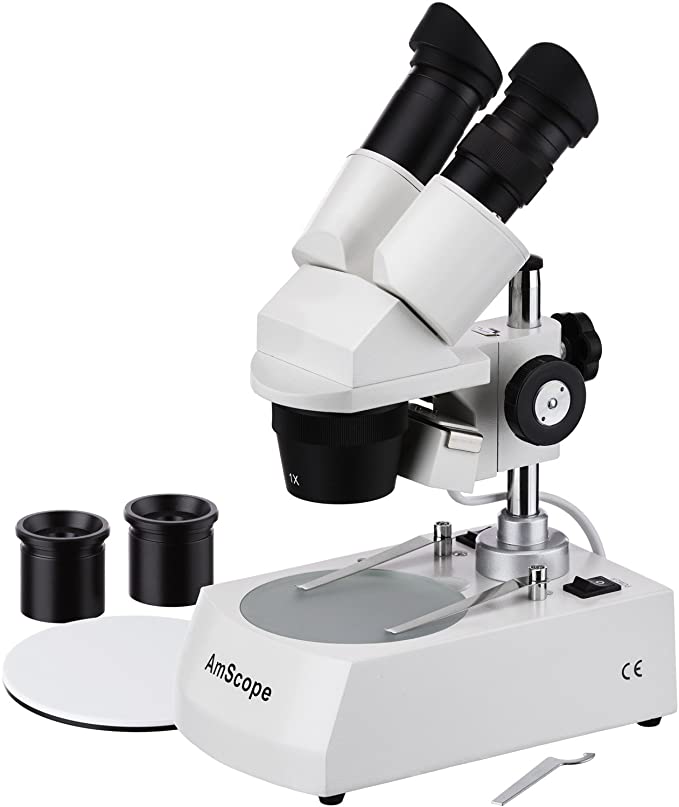
Dissecting microscope (Stereoscopic or Stereo microscope) This microscope is a dual-powered dissecting microscope of 10x-30x with an ability to rotate 360° making it ideal for viewing and focussing better to view samples. By rotating the lenses, users can change the magnification of image.

Dissecting microscope diagram with labels
Simple Microscope - Parts, Functions, Diagram and Labelling Simple Microscope - Parts, Functions, Diagram and Labelling A microscope is one of the commonly used equipment in a laboratory setting. A microscope is an optical instrument used to magnify an image of a tiny object; objects that are not visible to the human eyes. Table of Contents The common types of microscopes are: What is a Simple microscope? PDF Advanced Microscopy, Fall 2005 Week 1-Dissecting the Microscope 1. Draw a diagram of the polarizing microscope that you are using. Label all of the various parts of the scope. Make a sketch of the optical path of your microscope, locating the main parts (light source, objective, polarizer, analyzer, condensing lens, Bertrand lens, diaphragm, ocular (eye piece), stage, accessory slot, position of thin ... Parts of Dissecting Microscope | Botany - Biology Discussion Dissecting microscope is used to dissect small organisms or organs, e.g., embryo dissection. Its special utility is to observe such materials where high magnification is not needed. Design of Compound Microscope (With Diagram) | Biology Labelled Diagram of Compound Microscope
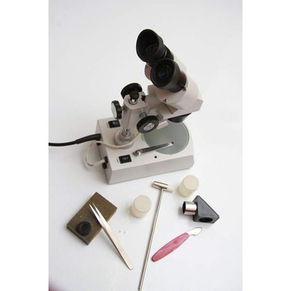
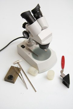
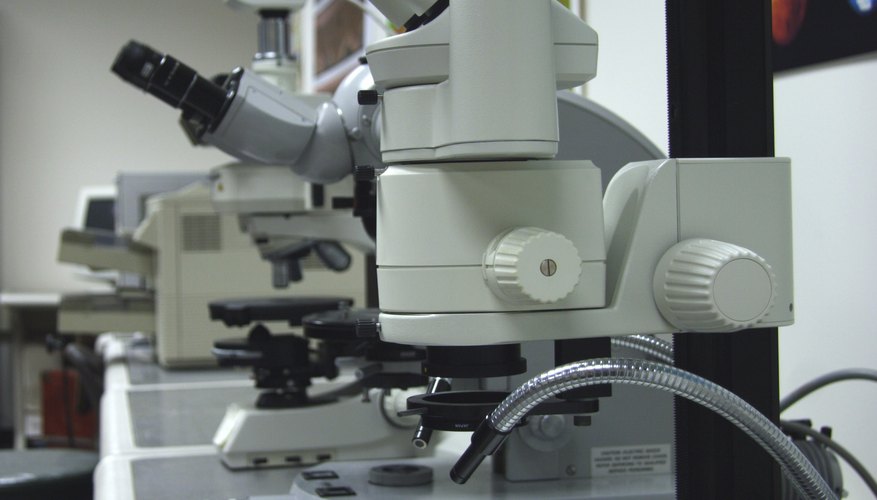
Dissecting microscope diagram with labels. Everything You Need to Know About A Dissecting Microscope A dissecting microscope, or more commonly known as a stereo microscope, is a microscope that gives a three-dimensional view of a specimen. This is because of the binocular head, or the two eyepieces that are slightly angled, which creates the perfect peripheral vision that results in a three-dimensional visual. Microscope Parts and Functions With Labeled Diagram and Functions How ... Body tube (Head): The body tube connects the eyepiece to the objective lenses. Arm: The arm connects the body tube to the base of the microscope. Coarse adjustment: Brings the specimen into general focus. Fine adjustment: Fine tunes the focus and increases the detail of the specimen. Solved Examine the sea urchin test under a dissecting | Chegg.com Transcribed image text: Examine the sea urchin test under a dissecting microscope. Find and label on the diagram the following: calcareous plates spine attachment sites tube feet perforations ambulacrum (the area where the tube feet from one radial canal exit the test) intra-ambulacral area Examine the Sea Biscuit and the Sand Dollar tests: • Describe where the tube feet are located and how ... Parts of Stereo Microscope (Dissecting microscope) - labeled diagram ... Stereo microscopes (also called Dissecting microscope) are branched out from other light microscopes for the application of viewing "3D" objects. These include substantial specimens, such as insects, feathers, leaves, rocks, sand grains, gems, coins, and stamps, etc. Functionally, a stereo microscope is like a powerful magnifying glass.
Compound Microscope Parts - Labeled Diagram and their Functions - Rs ... There are two major optical lens parts of a microscope: Eyepiece (10x) and Objective lenses (4x, 10x, 40x, 100x). Total magnification power is calculated by multiplying the magnification of the eyepiece and objective lens. The illuminator provides a source of light. The light is focused by the condenser and passing through the specimen placed ... Label the microscope Diagram | Quizlet Start studying Label the microscope. Learn vocabulary, terms, and more with flashcards, games, and other study tools. Labeling the Parts of the Microscope | Microscope World Resources Labeling the Parts of the Microscope This activity has been designed for use in homes and schools. Each microscope layout (both blank and the version with answers) are available as PDF downloads. You can view a more in-depth review of each part of the microscope here. Download the Label the Parts of the Microscope PDF printable version here. 16 Parts of a Compound Microscope: Diagrams and Video Once you have an understanding of the parts of the microscope it will be much easier to navigate around and begin observing your specimen, which is the fun part! The 16 core parts of a compound microscope are: Head (Body) Arm. Base. Eyepiece. Eyepiece tube.
Dissecting Microscope Parts - Like Hubble The dissecting microscope is also referred to as a stereoscopic microscope and is ordinarily used to study three-dimensional objects. And also as the name suggests for dissecting and analysing biological specimens under low magnification between two and two hundred and fifty times. Compound Microscope Parts, Functions, and Labeled Diagram Compound Microscope Definitions for Labels. Eyepiece (ocular lens) with or without Pointer: The part that is looked through at the top of the compound microscope. Eyepieces typically have a magnification between 5x & 30x. Monocular or Binocular Head: Structural support that holds & connects the eyepieces to the objective lenses. Parts of a microscope with functions and labeled diagram Figure: Diagram of parts of a microscope There are three structural parts of the microscope i.e. head, base, and arm. Head - This is also known as the body. It carries the optical parts in the upper part of the microscope. Base - It acts as microscopes support. It also carries microscopic illuminators. Microscope labeled diagram - SlideShare Microscope labeled diagram 1. The Microscope Image courtesy of: Microscopehelp.com Basic rules to using the microscope 1. You should always carry a microscope with two hands, one on the arm and the other under the base. 2. You should always start on the lowest power objective lens and should always leave the microscope on the low power lens ...
Compound vs Dissecting Microscope
Microscope, Microscope Parts, Labeled Diagram, and Functions Stage with Stage Clips: The stage of a microscope is a flat platform where you place your subject slides. Stage clips hold the slides in place. The mechanical stage of your microscope will help you to move the slide around by turning two knobs. One knobs moves it left and right, the other knobs moves it up and down.

Microscope World Blog: High School Microscope Features
Stereo Microscope Parts A stereo microscope has three key parts: Viewing Head/Body that houses the optical components in the upper part of the microscope. Focus Block that attaches the microscope head to the stand and focuses the microscope. Stand that supports the microscope and houses any integrated illumination. Stereo microscopes are increasingly modular.
Parts of the Dissecting Microscope | Synonym
Dissecting Stereo Microscope Parts and Functions Also known as a stereoscopic microscope, a dissecting microscope is a type of optical microscope commonly used for studying three-dimensional objects (3-D objects) as well as for dissecting biological specimen (e.g. insects and plant parts etc) at low magnification, between 2 and 100x depending on the microscope.

30 Label Parts Of Microscope - Labels Database 2020
Compound Light/Dissecting Microscope Diagram | Quizlet Used to examine material mounted on microscope slides (usually thinly sectioned & stained) Provides total magnification of 40x-1000x No space for dissection Rules TRANSPORT Arm & base USE Always start at 4x, Coarse focus, Fine focus Then change objectives & use fine focus as needed Coarse focus ONLY with 4x! CLEANING Objectives/Oculars

Microscope World Blog: November 2013
Labelled Diagram of Compound Microscope - Biology Discussion The below mentioned article provides a labelled diagram of compound microscope. Part # 1. The Stand: The stand is made up of a heavy foot which carries a curved inclinable limb or arm bearing the body tube. The foot is generally horse shoe-shaped structure (Fig. 2) which rests on table top or any other surface on which the microscope in kept.

33 Label The Microscope Quiz - Labels Design Ideas 2020
Label the microscope — Science Learning Hub Use this with the Microscope parts activity to help students identify and label the main parts of a microscope and then describe their functions. Drag and drop the text labels onto the microscope diagram. If you want to redo an answer, click on the box and the answer will go back to the top so you can move it to another box.
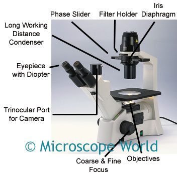
Microscope World Blog: Inverted Biological Microscopes
Binocular Microscope Anatomy - Parts and Functions with a Labeled Diagram Now, I will discuss the details anatomy of the light compound microscope with the labeled diagram. Why it is called binocular: because it has two ocular lenses or an eyepiece on the head that attaches to the objective lens, this ocular lens magnifies the image produced by the objective lens. Binocular microscope parts and functions
Parts of the Dissecting Microscope | Synonym
A Study of the Microscope and its Functions With a Labeled Diagram A Study of the Microscope and its Functions With a Labeled Diagram To better understand the structure and function of a microscope, we need to take a look at the labeled microscope diagrams of the compound and electron microscope. These diagrams clearly explain the functioning of the microscopes along with their respective parts.
Drawing Simple Dissecting Microscope Diagram - Micropedia
Parts of the Microscope with Labeling (also Free Printouts) 5. Knobs (fine and coarse) By adjusting the knob, you can adjust the focus of the microscope. The majority of the microscope models today have the knobs mounted on the same part of the device. Image 5: The circled parts of the microscope are the fine and coarse adjustment knobs. Picture Source: bp.blogspot.com.

Chapter 1 Questions PPT - BIOLOGY JUNCTION
Parts of the Dissecting Microscope - Synonym 6 Focus Knob The head of the microscope can be moved up and down with the focus knob, allowing the observer to view the image sharply; this is called rack and pinion focusing. 7 Stage Plate The specimen is placed on the stage plate for viewing. This plate is mounted on the base of the microscope, directly under the objective lens.

Compound Microscope Unlabeled - Micropedia
Parts of Dissecting Microscope | Botany - Biology Discussion Dissecting microscope is used to dissect small organisms or organs, e.g., embryo dissection. Its special utility is to observe such materials where high magnification is not needed. Design of Compound Microscope (With Diagram) | Biology Labelled Diagram of Compound Microscope
Difference Between Compound & Dissecting Microscopes | Sciencing
PDF Advanced Microscopy, Fall 2005 Week 1-Dissecting the Microscope 1. Draw a diagram of the polarizing microscope that you are using. Label all of the various parts of the scope. Make a sketch of the optical path of your microscope, locating the main parts (light source, objective, polarizer, analyzer, condensing lens, Bertrand lens, diaphragm, ocular (eye piece), stage, accessory slot, position of thin ...
36 Label The Parts Of A Microscope Worksheet - Labels Design Ideas 2021
Simple Microscope - Parts, Functions, Diagram and Labelling Simple Microscope - Parts, Functions, Diagram and Labelling A microscope is one of the commonly used equipment in a laboratory setting. A microscope is an optical instrument used to magnify an image of a tiny object; objects that are not visible to the human eyes. Table of Contents The common types of microscopes are: What is a Simple microscope?

All Saints Online

Dissecting Microscope With Labeled Parts - Micropedia

microscope labeled diagram simple microscope beauteous labeled - Top Label Maker
Homeschool Supplies & Science Supply List - Homeschool Den
Dissecting Microscopes - MicroscopeGenius.com
Post a Comment for "38 dissecting microscope diagram with labels"